The Marine Biological Association’s Microbiome Centre is home to unique and cutting-edge facilities which allow us to explore the cellular processes that shape the unique biology of marine life. Our world class microscopy equipment, including the Mesolens microscope, allow us to study marine organisms at cellular and subcellular resolution—and we’re proud to house one of only three Mesolens microscopes in existence!
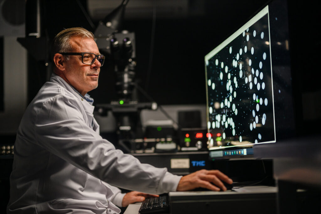
Dr Matthew Keys, Postdoctoral Researcher at the MBA, together with Professor Colin Brownlee, has been taking advantage of the wide field of view achieved by the Mesolens microscope to integrate chlorophyll fluorescence detection, measurement and visualisation for hundreds to thousands of phytoplankton cells simultaneously.
As well as forming the basis of marine food webs, microscopic phytoplankton in our ocean are responsible for 50% of global photosynthesis, which means they play a significant role in removal of carbon dioxide from the earth’s atmosphere while producing half of the oxygen that we breathe.
Chlorophyll is essential for photosynthesis, the process by which these organisms convert light energy into chemical energy. When chlorophyll, the green pigment found in all plants and phytoplankton, is illuminated it emits light (chlorophyll fluorescence). Over the past 25 years, measurement of changes in chlorophyll fluorescence under different light treatments has become one of the most popular techniques in plant and marine phytoplankton research to gain detailed information on the fundamental mechanisms of photosynthesis. This is usually conducted in closed chambers that provide a single collective value for a whole population.
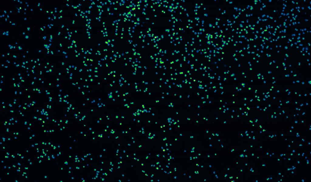
However, more recently, quantitative imaging of chlorophyll fluorescence has emerged, which enables us to measure and visualise important parameters of photosynthesis within single cells. As phytoplankton are microscopic in size, current commercial systems are designed around conventional microscopes which have a small field of view – 0.5 mm. That means we can only measure and visualise chlorophyll fluorescence in a small number of cells with these systems. In contrast, the Mesolens microscope has a 5 mm field of view (25 mm2) which means we can visualise hundreds to thousands of cells in a single field of view.
We have now integrated chlorophyll fluorescence detection techniques with the Mesolens microscope by combining state-of-the-art electronics, powerful LED lights and computer software.
These new capabilities mean we can analyse photosynthetic performance of each cell individually in natural phytoplankton populations from seawater samples. This is an important advancement as we can now understand, for example, which phytoplankton species respond to changing environmental conditions within natural populations.
Preliminary analyses using the Mesolens chlorophyll fluorescence imaging system have highlighted a very wide range of single cell photosynthetic performance within natural populations, enabling us to drill down to a much finer level of detail than was previously possible.
More broadly, the Mesolens is being utilised at the MBA to study a wide range of physiological processes in populations or multicellular structures that are not amenable to conventional light microscopy.
These range from assessing the signalling processes that allow different phytoplankton cells in a population to respond to environmental cues, to understanding how the opening and closing of every plant stomata, the tiny pores on the surface of a plant leaf, is coordinated.
Discover our research here.